Clinical ECG Interpretation
iOS Universel / Medecine
Many medical students in their clinical years have a good level of competency in interpreting the primary ECG parameters, but their ability to recognize ECG signs of emergencies and common heart abnormalities is usually low. ECG interpretation skills are determined not only by attendance at regular ECG classes but by self-education as well. Electrocardiogram (ECG) interpretation is an essential skill for emergency medicine (EM) physicians.
Electrocardiography is the process of producing an electrocardiogram (ECG or EKG), a recording of the heart's electrical activity through repeated cardiac cycles. It is an electrogram of the heart which is a graph of voltage versus time of the electrical activity of the heart using electrodes placed on the skin. These electrodes detect the small electrical changes that are a consequence of cardiac muscle depolarization followed by repolarization during each cardiac cycle (heartbeat).
During each heartbeat, a healthy heart has an orderly progression of depolarization that starts with pacemaker cells in the sinoatrial node, spreads throughout the atrium, and passes through the atrioventricular node down into the bundle of His and into the Purkinje fibers, spreading down and to the left throughout the ventricles. This orderly pattern of depolarization gives rise to the characteristic ECG tracing. To the trained clinician, an ECG conveys a large amount of information about the structure of the heart and the function of its electrical conduction system. Among other things, an ECG can be used to measure the rate and rhythm of heartbeats, the size and position of the heart chambers, the presence of any damage to the heart's muscle cells or conduction system, the effects of heart drugs, and the function of implanted pacemakers.
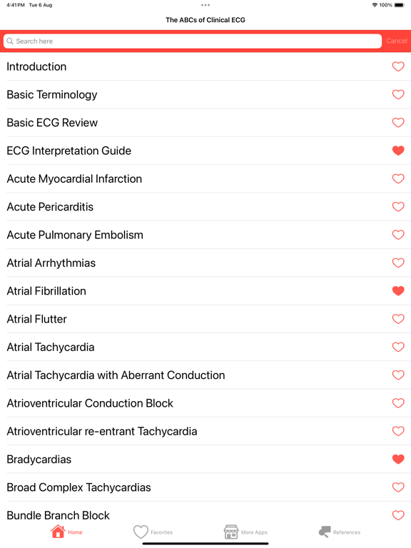
App content:
1. Introduction
ECG history
Leads
Heart Rate
Rhythm
Cardiac Axis
Relationship Between Heart Rate, Rhythm and Axis
Signal Processing In ECG
ECG Paper Study and interpretation
2. Basic Terminology
P Wave and PR Interval
QRS Complex
ST Segment
T Wave
QT Intervals
U Wave
ECG Terminology Review
3. Basic ECG Review
MI & ECG
Thyroid Disorder And ECG
Arrhythmia and ECG
Emergency ECG
4. ECG Interpretation Guide
ECG Rules
Approach to Interpretation of ECG
Commenting on ECG
5. Acute Myocardial Infarction
6. Acute Pericarditis
7. Acute Pulmonary Embolism
8. Atrial Arrhythmias
9. Atrial Fibrillation
10. Atrial Flutter
11. Atrial Tachycardia
12. Atrial Tachycardia with Aberrant Conduction
13. Atrioventricular Conduction Block
14. Atrioventricular re-entrant Tachycardia
15. Bradycardias
16. Broad Complex Tachycardias
17. Bundle Branch Block
18. Cardiomyopathies
19. Chronic Obstructive Pulmonary Disease
20. Fascicular Blocks
21. Heart Block
22. Hypercalcaemia
23. Hyperkalaemia
24. Hypokalaemia
25. Hypothermia
26. Hypothyroidism
27. Junctional Tachycardias
28. Normal ECG and Variation with Respiration
29. Polymorphic Ventricular Tachycardia
30. Posterior Myocardial Infarction
31. Right Atrial Enlargement
32. Right Sided Valvular
33. Right Ventricular Hypertrophy
34. Right Ventricular Infarction
35. Right Ventricular Outflow Tract Tachycardia
36. Thyrotoxicosis
37. Ventricular Tachycardias
38. ecg cases with answers
Supports dark mode & favoriting
Care has been taken to provide accurate information on this app in keeping with current ecg teaching. It is intended only for use by individuals who are students or medical practitioners in the field. It is not intended to replace formal medical ecg training. It is not intended for use by non-medical personnel. This information is provided for general medical education purposes only and is not meant to substitute for the independent medical judgement of a physician relative to diagnostic and treatment options of a specific patient's medical condition. In no event will the authors or editors or the developer of this app be liable for any decision made or action taken in reliance upon the information provided through this application.
Quoi de neuf dans la dernière version ?
What is difference between EKG and ECG?
So what's the difference? EKG and ECG are actually different spellings of the same diagnostic test that monitors your heart's electrical activity. EKG is the abbreviation from the German spelling of electrocardiogram (which is elektrokardiogramm in German).
How does EKG work?
Electrocardiogram | Johns Hopkins Medicine
Electrodes (small, plastic patches that stick to the skin) are placed at certain spots on the chest, arms, and legs. The electrodes are connected to an ECG machine by lead wires. The electrical activity of the heart is then measured, interpreted, and printed out. No electricity is sent into the body.


